Revision as of 22:07, 15 March 2013 by
JKim (Talk)
Introduction
Macroscopic Entries
Microscopic Entries
| Epidermis of the thallus showing highly contoured surface and pink glandular material of Palmaria palmata when viewed at 400x with Acidified Chloral Hydrate Glycerol Solution.cellular structures identified in Palmaria palmata are the epidermis of the thallus showing highly contoured surface and pink glandular material and parenchyma cells of the thallus showing pink pigmentation when viewed at 400x with Acidified Chloral Hydrate Glycerol Solution.
Source: Elan M. Sudberg, Alkemist Laboratories [1]
|
|
|
|
HPTLC Entries
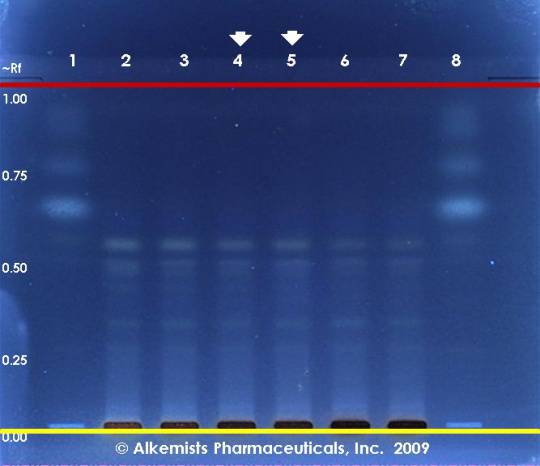
Palmaria palmata HPTLC ID - 10% Ethanolic H2SO4 ->115° C 15 min -> UV 365 nm
Dulse (seaweed) (Palmaria palmata)
Lane Assignments Lanes, from left to right (Track, Volume, Sample):
- 15 μL Fucoxanthin ~0.1% in CH3OH
- 5 μL Palmaria palmata-1 (seaweed)
- 5 μL Palmaria palmata-2 (seaweed)
- 5 μL Palmaria palmata-3 (seaweed)
- 5 μL Palmaria palmata-3 (seaweed)
- 5 μL Palmaria palmata-4 (seaweed)
- 5 μL Palmaria palmata-5 (seaweed)
- 15 μL Fucoxanthin ~0.1% in CH3OH
Reference materials used here have been authenticated by macroscopic, microscopic &/or TLC studies according to the reference source cited below held at Alkemists Laboratories, Costa Mesa, CA.
Stationary Phase Silica gel 60, F254, 10 x 10 cm HPTLC plates
Mobile Phase cyclohexane: ethyl acetate: propionic acid [6/3.8/0.2]
Sample Preparation Method 0.3 g + 3 ml 80% CH3OH sonicated + heated @ 50° C ~ 1 hr
Detection Method 10% Ethanolic H2SO4 ->115° C 15 min -> UV 365 nm
Reference see Adapted from Pharmacognosy Phytochemistry Medicinal Plants, Bruneton, J. 1995
Source: Elan M. Sudberg, Alkemist Laboratories [2]
|
Other Points of Interest
Cite error: <ref> tags exist, but no <references/> tag was found
_Kuntze_-Palmariaceae--1.jpg)
